Oral surgery
Jaw Cyst Surgery in Istanbul
Medically reviewed by Dt. Mehmet Alp Erdem·Last updated:

What is a jaw cyst?
A cyst is a small sac lined by a thin membrane and filled with fluid or soft material. Most cysts in the jaw are odontogenic, meaning they arise from tissues involved in tooth development or from inflammation around a tooth. They tend to grow slowly and can stay completely silent for years.
That is why many jaw cysts are discovered by chance on an X-ray taken for something else. Being told there is a cyst in your jaw can be unsettling, but it helps to know that, according to sources such as Cleveland Clinic, most jaw growths are benign and malignant ones are rare. The aim is simply to identify the type and treat it before it grows.
Common types of jaw cyst
The type of cyst guides both the surgical approach and how long it needs to be followed up. Imaging gives strong clues, but the final diagnosis comes from examining the removed tissue under a microscope. The types seen most often are:
- Radicular (periapical) cyst: forms at the root tip of a tooth whose nerve has died; the most common type.
- Dentigerous cyst: develops around the crown of an unerupted tooth, very often a wisdom tooth.
- Odontogenic keratocyst: less common, with a higher tendency to recur, so it is followed up for longer.
- Benign odontogenic tumours: lesions such as odontomas or ameloblastomas are uncommon and are planned individually.
| Type | Where it forms | Recurrence and follow-up |
|---|---|---|
| Radicular (periapical) cyst | At the root tip of a tooth whose nerve has died | Usually does not return once fully removed |
| Dentigerous cyst | Around the crown of an unerupted tooth, often a wisdom tooth | Usually does not return once fully removed |
| Odontogenic keratocyst | Less common | Higher tendency to recur; regular X-rays for several years |
| Benign odontogenic tumours | Uncommon lesions such as odontomas or ameloblastomas | Planned individually |
Symptoms and diagnosis
Small cysts rarely cause symptoms. As they enlarge, you might notice a slow-growing swelling of the jaw that often doesn't hurt, teeth shifting or becoming loose, a denture that no longer fits, or pain and discharge if the cyst becomes infected. Many of these signs have other, simpler causes, so assessment always begins with an examination.
Dt. Mehmet Alp Erdem, a dentist focused on oral surgery and a PhD candidate in Oral and Maxillofacial Surgery at Yeditepe University, uses a 3D cone beam CT (CBCT) scan alongside the panoramic X-ray. The scan shows the cyst's borders and its relationship to tooth roots, the jaw nerve and the sinus. Pulp vitality tests help decide whether the associated tooth can be saved.
How jaw cyst surgery is done
Most cysts are treated by enucleation: the whole cyst, including its lining, is removed in one piece. This is normally done under local anaesthetic. A small flap of gum is lifted, the cyst is reached through the bone, removed, and the area is stitched.
A related tooth may be kept with root canal treatment and, if needed, an apicoectomy (root-end surgery), or it may need to be extracted.
For very large cysts close to important structures, a staged approach is often safer. Marsupialisation or decompression drains the cyst and keeps a small opening so that it gradually shrinks; the remaining lining is removed in a second, smaller procedure. Where a large defect is left and an implant is planned later, a bone graft may be considered.
In every case, the removed tissue is sent for histopathology. Imaging offers strong clues about a lesion, but only the laboratory report confirms exactly what it was. That report sets the follow-up schedule and shows whether anything further is needed, and it is talked through with you at your review appointment.
After removal: bone, teeth and implants
Once a cyst is removed, the cavity it leaves usually fills with your own new bone over time, and small to medium defects often need no added material. A bone graft is considered when the cavity is large, the jaw has become thin, or an implant is planned in that area later.
If a related tooth was kept, a sound root canal filling and a well-sealed crown or filling are important for preventing new inflammation. If a tooth was removed, the gap can be restored with an implant, a bridge or another option once a follow-up scan shows the bone has healed well enough.
The underlying principle is to preserve healthy tissue and avoid unnecessary intervention. Each step is planned on the result of the previous one, so treatment is completed with the least intervention that does the job properly.
Recovery, risks and follow-up
Some swelling and tenderness are expected for a few days, and most people return to work within two to four days. The bone itself takes longer: depending on the size of the cavity, it can fill in over several months to around a year, during which you should avoid putting heavy force on the area.
Possible complications include infection, temporary sensitivity in neighbouring teeth and, for lower-jaw cysts near the nerve, temporary lip numbness; CBCT planning is designed to reduce these risks. Radicular and dentigerous cysts usually do not return once fully removed, while types such as odontogenic keratocysts need regular X-ray checks for several years.
How treatment works
- 01
Examination and CBCT
A panoramic X-ray and 3D scan show the size and borders of the cyst and nearby structures.
- 02
Treatment plan
The approach, what happens to the related tooth, any bone graft and the recovery are explained in writing.
- 03
Surgery
The cyst is usually removed in one piece under local anaesthetic; very large cysts may first be decompressed.
- 04
Histopathology
The tissue is analysed in a laboratory to confirm the diagnosis and guide follow-up.
- 05
Follow-up
Bone healing and any sign of recurrence are checked with X-rays every 6-12 months as needed.
Travelling to Istanbul for jaw cyst surgery
If you already have a panoramic X-ray or CBCT scan, you can send it via WhatsApp for a preliminary review. It gives a sense of the cyst's size and the likely approach before you plan travel; the final plan follows the in-person examination.
For most cysts one trip to Istanbul is enough, and staying around 7-10 days allows the stitch check before you fly home. The histopathology report can be sent to you digitally, and long-term follow-up X-rays can be taken by your local dentist and shared remotely. Very large cysts treated in stages may need a second visit.
Frequently asked questions
Is a jaw cyst serious?
Most jaw cysts are benign and very treatable. Left alone, though, a cyst can slowly expand, thin the jawbone, move teeth or become infected. That is why a cyst seen on an X-ray is usually assessed with a 3D scan and, in most cases, removed and analysed.
Can a jaw cyst go away on its own?
True jaw cysts rarely disappear on their own. Some small inflammatory lesions at a root tip do heal after successful root canal treatment, but an established cyst usually needs to be removed. Your X-rays and CBCT scan help show which situation applies to you.
Is jaw cyst surgery done under general anaesthetic?
Most jaw cysts can be removed under local anaesthetic in the dental chair. Very large lesions that involve much of the jaw, or sit close to major structures, may need general anaesthetic in a hospital setting. This is decided after the CBCT assessment.
Will I lose a tooth because of the cyst?
Not necessarily. A tooth linked to a radicular cyst can often be saved with root canal treatment and, if needed, an apicoectomy. If the tooth is badly damaged, or the cyst surrounds an impacted wisdom tooth, extraction may be advised. This is discussed in detail before surgery.
Why is the tissue sent to a laboratory?
X-rays and scans show the shape of a lesion but cannot confirm what it is. Histopathology, the microscopic examination of the removed tissue, gives a definite diagnosis and decides how long you need follow-up. Results usually arrive within one to two weeks and can be shared digitally.
Can a jaw cyst come back after surgery?
Radicular and dentigerous cysts usually do not recur after complete removal. Some types, particularly odontogenic keratocysts, have a higher recurrence tendency, so they are followed with periodic X-rays for several years. Your follow-up schedule is based on the laboratory report.
Can I have a dental implant where the cyst was?
In most cases, yes, once the bone has healed. If the cavity is large, a bone graft may be placed at the time of surgery or later. Implant timing is decided when a follow-up scan shows enough new bone in the area.
How long does recovery from jaw cyst surgery take?
The gum usually heals in two to three weeks, and most people are back at work within a few days. The bone cavity takes longer, from several months to around a year depending on its size, and its progress is checked with follow-up X-rays.
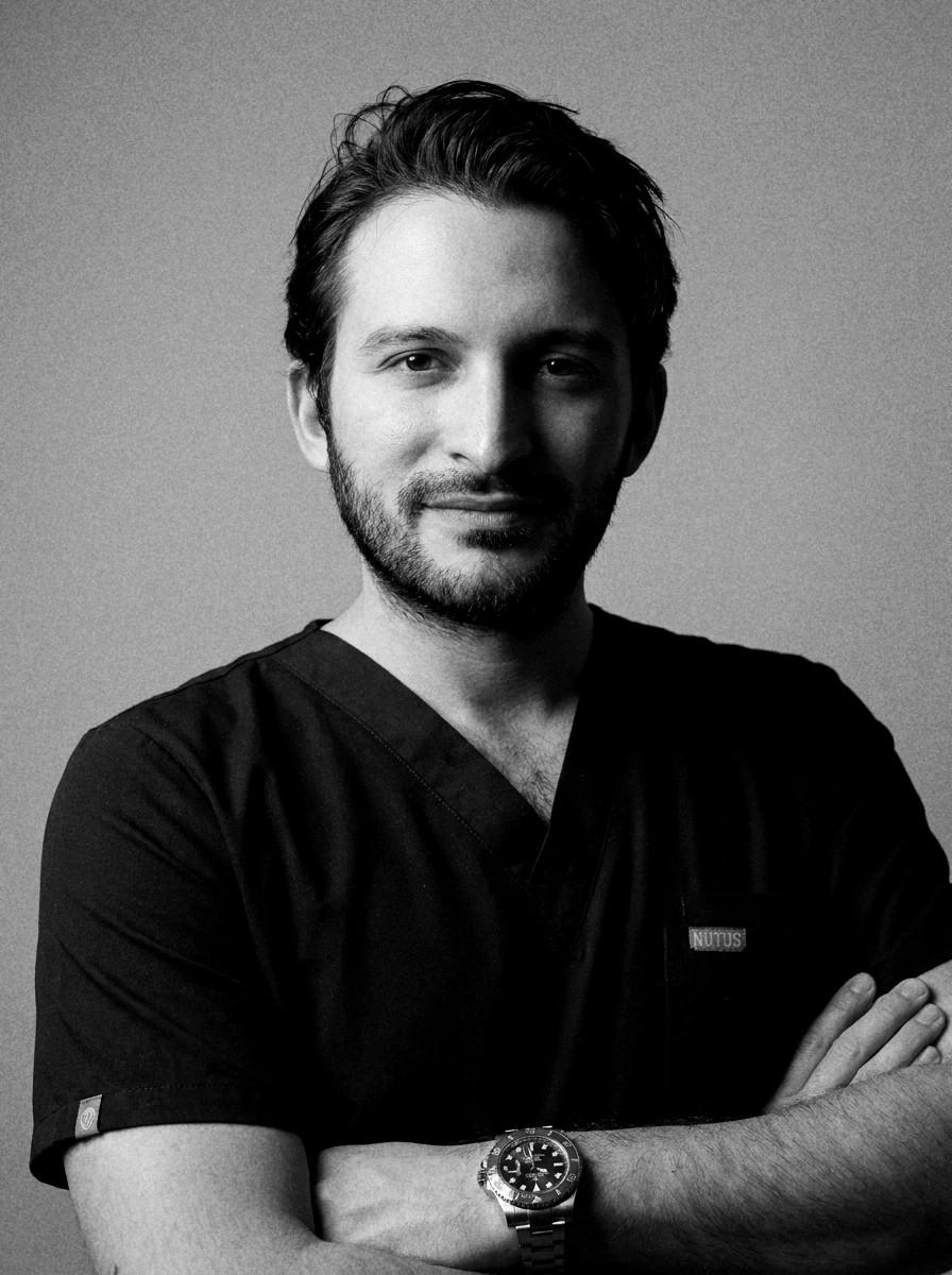
Written and reviewed by
Dt. Mehmet Alp ErdemDentist · PhD candidate in Oral and Maxillofacial Surgery, Yeditepe University
Sources
This page is for general information. Diagnosis and treatment decisions are made by the dentist after examination and imaging.
Plan your consultation
If you already have X-rays or a CBCT scan, send them on WhatsApp for a preliminary review before you travel.
Related treatments

Wisdom Tooth Removal in Istanbul
Planned removal of impacted or partly erupted wisdom teeth, guided by 3D CBCT imaging and focused on protecting surrounding tissue.
Details
Root Canal Treatment in Istanbul
Treatment that removes inflamed or infected pulp, cleans and seals the root canals and aims to keep your natural tooth.
Details
Dental Bone Grafting in Istanbul
Rebuilding lost jawbone so implants have a stable foundation — graft types, when grafting is needed, healing time and trips to Istanbul.
Details
Dental Implants in Istanbul
A titanium or zirconia root placed in the jawbone to replace a missing tooth — planning, healing time, trips to Istanbul and aftercare.
Details